BioFrontiers Core Facility Instruments
BioFrontiers Core Facility Instruments
Please inquire regarding the following:
Cell Culture Equipment: Biosafety Hoods, incubators, centrifuges, liquid nitrogen storage
Analysis Equipment: spectrophotometer, fluorimeter, calorimeter
Note: Adequate experience and/or training is required for individual usage as well as current lab safety training credentials. Biofrontiers personnel are happy to help with consultation and initial setup without charge. Biofrontiers reserves the right to observe and monitor all procedures.
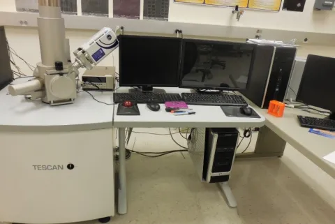
Scanning Electron Microscope -Model: TESCAN VEGA3 SBU
Fully integrated with a selected energy dispersive X-ray microanalyser (EDX) for chemical and elemental analysis
Technical characteristics:
- Resolution: about 5 nm
- Magnification: up to 300,000x
- Energy Dispersive X-ray microanalysis (EDXA) capability. Can measure elemental surface composition with about 1% accuracy.
- Can change samples in under 1 minute
Sputtering/Etch Unit Denton Vacuum Desk V (HP)
Deposits a thin conductive coating on a sample to avoid charging effects in the SEM
The thickness of the deposited layer can be controlled by changing technical parameters (current, time, type of the target etc.).
An example of the performance for target material gold in air: Target material: gold. Gas: air Pressure: 50 mTorr
Time: 600 s
Power | Current | Film Thickness | Deposition Rate |
|---|---|---|---|
% | mA | Å | Å/s |
38 | 10 | 260 | 0.43 |
58 | 20 | 650 | 1.08 |
Electron Beam Lithography (NPGS - Nanometer Pattern Generation System).
By Using the SEM Vega/NPGS, - Can write patterns in photoresist with a resolution of about 40 nm. Detailed information on the NPGS can be found at http://www.jcnabity.com/
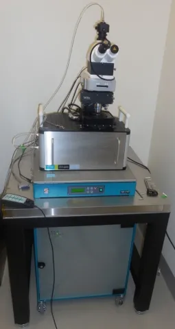
Scanning Near-Field Optical Microscope WITec alpha300 S including Acoustic AC Mode
User-Friendly
Scanning Near-field Optical Microscopy (SNOM) modes; Atomic Force Microscopy (AFM) modes
Standard resolution - less than 90 nm
Research grade optical microscope with 6 x objective turret
· Video system: eyepiece color video camera
· LED white-light source for Köhler illumination of tip and sample
· High sensitivity b/w video camera to view sample and SNOM/AFM tip in transmission
· Manual sample positioning in x- and y-direction, 25 mm travel
· Microscope base with active vibration isolation system
· Piezo-driven scan stage (scan range 100 x 100 x 20 µm; others optional)
Sample Size:
Usually 120 mm in x- and y-direction, 25 mm in height
Scanning Near-field Optical Microscopy (SNOM) mode; Atomic Force Microscopy (AFM) mode
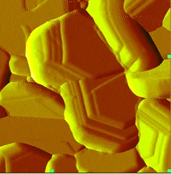
Atomic Force Microscope/Magnetic Force Microscope
Veeco di MultiMode V
A scanning probe system where a sharp tip, attached to a cantilever, is moved across the surface of a sample.
Forces on the tip cause bending of the cantilever which is monitored by a laser beam.
Features:
· Vertical resolution 1 nm
· Horizontal resolution 5 nm
· Imaging modes - Contact mode, tapping mode, non-contact mode
· Small sample scanning (less than 15 mm diameter, 8 mm thickness)
TCS SP5 Confocal Microscope - Leica
The SP 5 laser scanning confocal microscope uses laser excitation for fluorescent and transmitted light measurements. There are three PMTs to collect fluorescent emissions. Another PMT can be used to collect transmitted information. The fluorescence collection system allows the filters to be changed in bandwidth and wave length from 360-780 nm.
A motorized stage in x, y, and z allows relocation acquisitions and 3 d reconstructions to be done.
Horizontal Resolution - about 180 nm
Vertical Resolution - about 550 nm
Up to 7 laser excitation wavelengths can be used: 405 nm, 456 nm, 476 nm, 488 nm, 514 nm, 532 nm, 633 nm.
Objectives
63x 1.4-0.60 na oil ~ 40x 1.3 na oil ~ 20x 0.70 na air ~ 10x 0.25 na air
Visual Filter
Ex Dm Em A cube BP340-380 400 LP 425
I3 cube BP 450-490 510 LP 515
N2.1 cube BP 515-560 580 LP 590
Lasers
Diode 405 nm ~ Argon 458, 476, 488, 514 nm ~ DPSS 561 nm ~ HeNe 633 nm
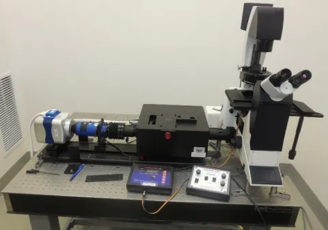
TIRF (Total Internal Reflection Fluorescence) Microscope
A total internal reflection fluorescence microscope (TIRFM) is a type of microscope with which a thin region of a specimen, usually less than 200 nm can be observed. TIRFM is an elegant optical technique utilized to observe single molecule fluorescence at surfaces and interfaces. The technique is commonly employed to investigate the interaction of molecules with surfaces, an area which is of fundamental importance to a wide spectrum of disciplines in cell and molecular biology.
Features:
· DMI3000 B microscope
· Objective: Leica HCX PL APO 100x/1.47. An objective heater controller included.
· Polarization control (Meadowlark optics)
· 16 x16 µm EMCCD Camera
Laser lines: 405 nm (100 mW);
480 nm (50 mW); 560 nm (50 mW)
Requires training and monitoring from BioFrontiers staff
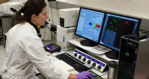
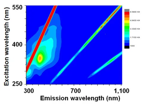
Fluorescence spectrometer Nano Log Horiba iHR320
Features
- Frequency range - Visible to near IR
- A complete excitation-emission spectrum scan is taken in seconds
- Excitation wavelengths 250-550 nm
- Emission wavelengths 300-1,300 nm
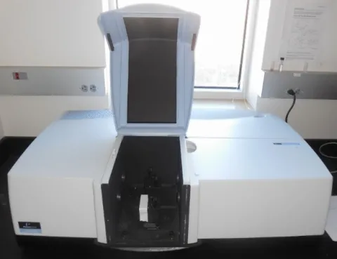
Spectrophotometer (UV - Visible - near IR)
A double beam (sample + control), double monochromator, ratio recording UV/Vis/NIR. Good sensitivity, resolution and speed in the NIR range, it simplifies the analysis of difficult samples such as high absorbing glass, optical coatings or thin film filters.
- Spectral range: 175 nm - 3300 nm.
Wavelength Resolution:
- Better than 0.05 nm (UV-Visible);
- Better than 0.20 nm (NIR).
Low-field NMR Spectrometer
- Low magnetic field (364 mT) means no cryogenic cooling is necessary for the magnet
- Sample temperatures can be varied from 5 oC to 60 oC
FC500 Flow Cytometer - Beckman/Coulter
Flow cytometers are used to rapidly analyze particles one at a time which allows the identification of numerous subpopulations a statistically significant numbers. Flow rates of 3000 particles per second are possible on this instrument. Cells are the usual particles analyzed. Up to 24 parameters can be measured; 16 parameters simultaneously. Particle size range from 0.5 to 40 microns.
These parameters can be measured on either a linear or a 4 decade log scale. Normally the parameters are forward scatter (FS), side scatter (SS), and five fluorescent wavelengths at 525, 575, 620, 675, and 755 nm. Two laser excitation wavelengths are available 488 nm and 635 nm. The lasers are coaxial. Additional parameters are peak, time, and ratio. Samples may be run in 12x75 mm tube, 96 well plates or 24 well plates.
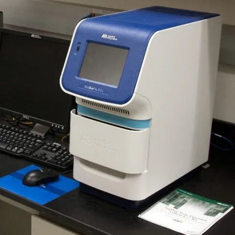
StepOne Plus Real-Time PCR - Applied Biosystems
The system can be used to detect and quantify DNA and other real-time PCR applications.
This instrument uses a 96 well plate and 4 colors.
The use of this instrument will require the user to provide assays, reagents and other materials for the test desired - we can assist with the ordering of necessary materials.
Detailed information on the RealTime PCR machine can be found at: http://tools.invitrogen.com/content/sfs/brochures/cms_042763.pdf
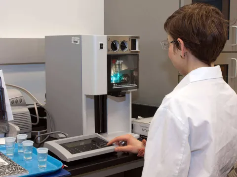
Coulter Counter Z2
The Z2 counts particles passing through an orifice based on the change of electrical resistance in the orifice. This allows not only counting of the particles but also allows sizing based on the resistance change.
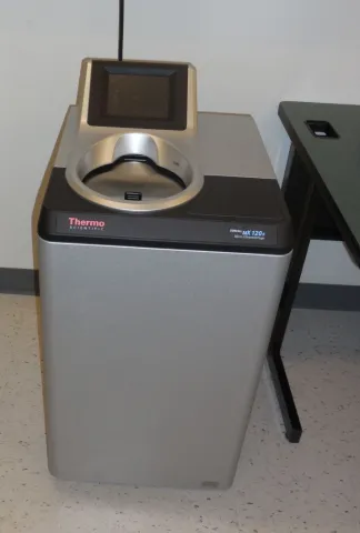
Ultracentrifuge Model: Sorvall MX 120 Plus Micro-Ultracentrifuge
Separates liquid-suspended materials having different densities and particle size.
Features
- The maximum speed is 120,000 rpm (649,826 xg)
- S120-AT2 Rotor - capacity: 10x2.0 ml, maximum speed 120,000 rpm
- S50-ST Rotor (capacity: 4x7.0 ml, maximum speed 50,000 rpm
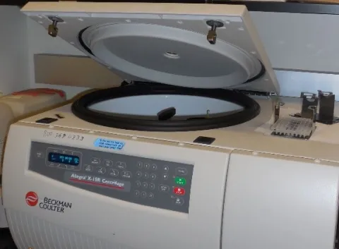
S50-ST Rotor (capacity: 4x7.0 ml, maximum speed 50,000 rpm Centrifuge Model: Allegra X-15R centrifuge
- A refrigerated bench-top centrifuge
- Applications include pelleting, extractions, purifications, phase separations, receptor binding, column centrifugations, cell isolation.
- Rapid sedimentation of protein precipitates, large particles, and cell debris.
- Available rotors: SX4750 swinging bucket rotor (tube-and bottle (50 ml x 5 or 15 ml x 19) buckets and multiwall-plate carriers, maximum speed is 4450 rpm (4060 xg)).
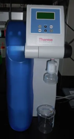
Water purification system Model: MicroPure UV/UF
A source of the ultra-purified water
Features
- 18.2 MΩ·cm at 25 0C
- At the flow rate 1 l/min